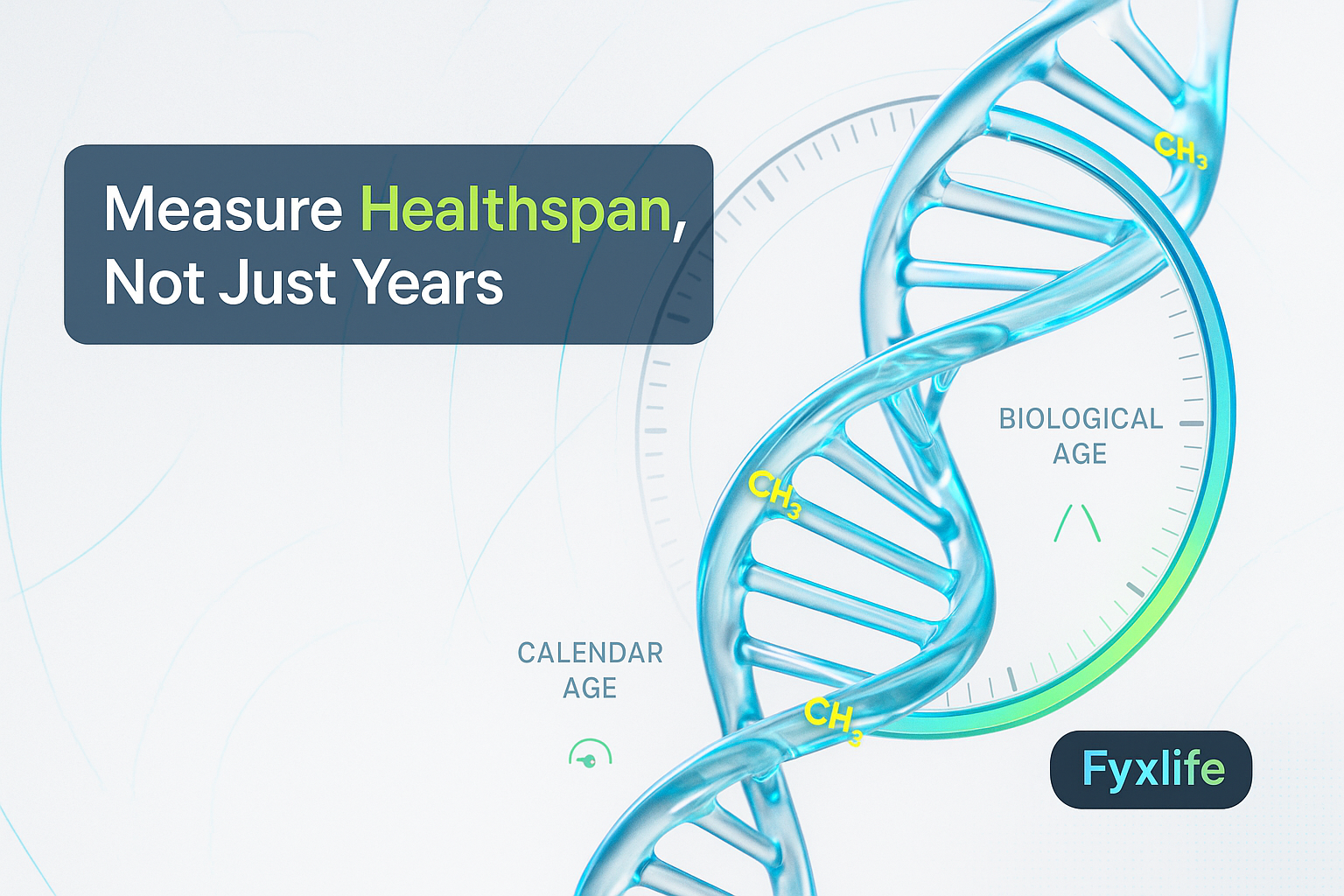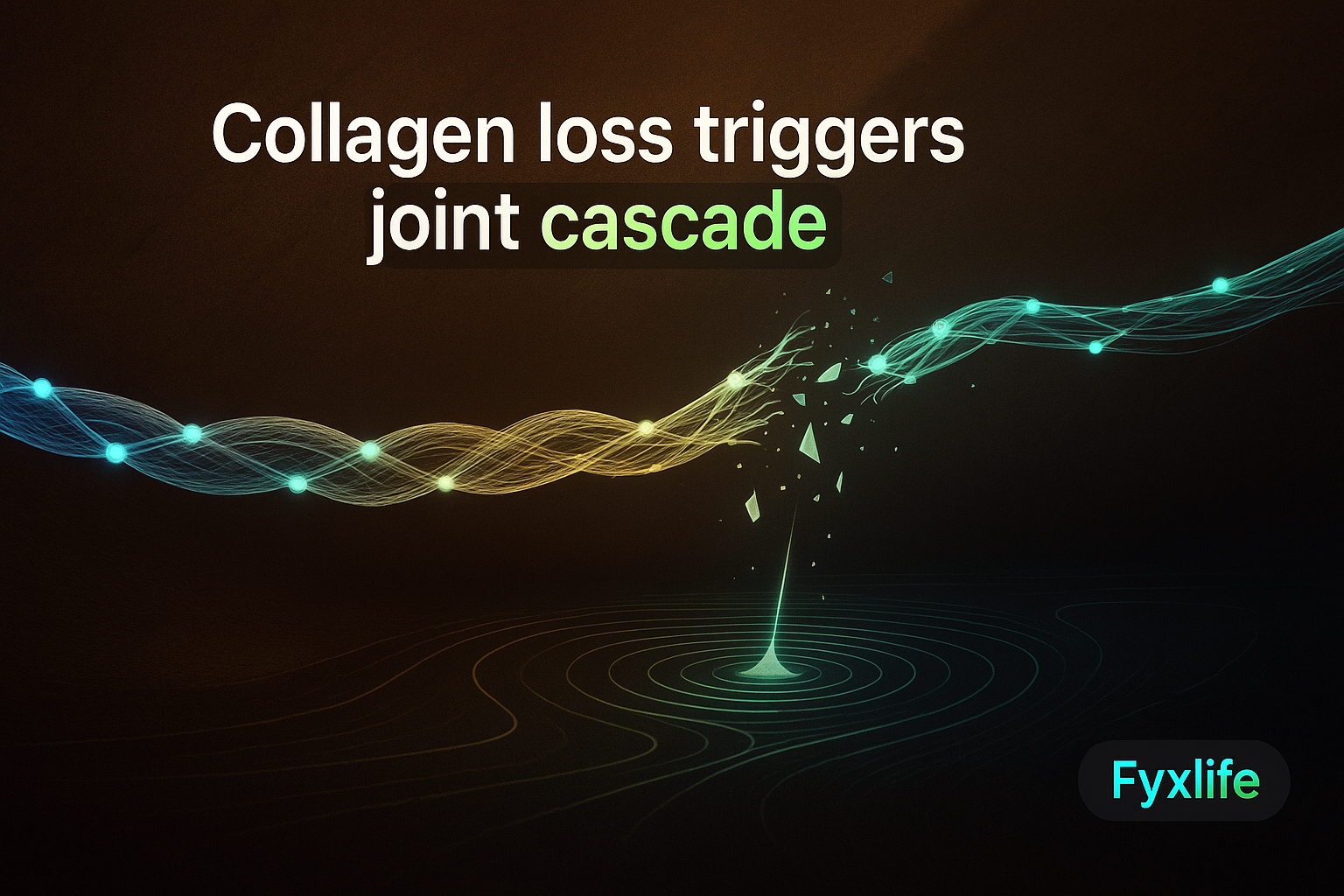
You don’t feel your cartilage breaking down — until the morning stiffness arrives, then the inflammation, then the pain that won’t settle. What looks like a joint problem in your knee is often the final stage of a cascade that started years earlier, quietly, with collagen loss you never noticed. By the time it hurts, the biology has already moved through several stages. Understanding those stages changes how you think about every supplement on the shelf.
Your Joint Isn’t the Problem — It’s the Last Domino
What collagen actually does inside a joint (and why it’s irreplaceable)
Most people think of collagen as a beauty supplement. That framing undersells it dramatically. Inside a joint, collagen is structural load-bearing architecture. Specifically, it is the primary component of the extracellular matrix (ECM) — the dense scaffolding that surrounds and supports your joint cells, giving cartilage both its compressive strength and its ability to absorb and distribute force across every step you take. Collagen plays a critical role in maintaining the mechanical properties of articular cartilage and the stability of this matrix — and many pathogenic processes in joint disease directly involve disruption of this collagen framework.
The dominant collagen in healthy cartilage is type II collagen — a tightly coiled, resilient protein that forms interlocking fibre networks capable of withstanding years of repeated mechanical load. It is not easily replaced. And unlike bone, which remodels constantly, cartilage has almost no blood supply, which means its repair capacity is genuinely limited. When type II collagen fibres degrade, the joint cannot simply rebuild them on demand.
The slow drain: how collagen loss begins before symptoms appear
Think of your joint cartilage as a suspension bridge. Collagen fibres are the cables — under constant load, slowly fraying. You don’t notice one fraying cable. You probably don’t notice ten. The bridge continues to function. But once enough cables snap, the deck cracks, debris falls into the water below, the river authority — your immune system — sends an emergency response team, that team triggers a secondary flood-control protocol, and suddenly you have a multi-agency crisis that started with a single fraying cable years ago.
This is not a metaphor for dramatic effect. It is an accurate description of the biology. The collagen degradation phase is silent because your body has redundancy built in. Pain receptors in cartilage are sparse. The ECM absorbs insult quietly until a threshold is crossed. By the time your knee aches getting out of a chair, you are likely well past stage one of the cascade.
Stage 1 — Structural Failure: When Type II Becomes Type I
Healthy cartilage collagen vs scar-tissue collagen — why the swap matters
The first measurable sign that something has shifted inside a deteriorating joint is a change in the type of collagen being produced. In a healthy joint, specialised cartilage cells called chondrocytes manufacture type II collagen — the high-performance version. Under sustained stress or degradation, these cells begin producing type I collagen instead. Type I is the body’s workhorse repair collagen — the scar-tissue version. It is what fills in a wound. It is structurally inferior for cartilage under compressive load.
In osteoarthritic knee cartilage, type I collagen is upregulated — a marker that normal cartilage collagen is being replaced by a fibroblast-like version, signalling failed repair rather than regeneration. This is a critical distinction. The joint is still trying to repair itself. But it has reached for the wrong tool — the biological equivalent of patching a load-bearing steel cable with duct tape.
What this shift signals about the state of your joint repair system
The type I collagen swap is not just a local cartilage problem. Collagen biomaterials regulate the signalling mechanisms of bone and immune cells involved in tissue repair — meaning any imbalance in collagen disrupts these pathways systemically, with consequences extending well beyond the local joint. When your body’s collagen architecture is compromised, the disruption propagates. Bone remodelling signals change. Immune cell behaviour changes. The joint is deteriorating, but it is also sending distress signals upstream that alter how your entire musculoskeletal system operates.
This is also where the cascade becomes difficult to reverse. Multiple signalling pathways govern the cellular and molecular processes of cartilage repair — and once those pathways have shifted into damage-control mode, simply adding collagen from outside the body faces an uphill battle against biological momentum.
Stage 2 — The Immune Alarm: Complement Activation and the Inflammatory Flood
How cartilage debris activates your immune system’s amplification loop
When cartilage breaks down, it does not just disappear. Collagen fragments, proteoglycan debris, and cellular breakdown products spill into the joint space. Your immune system registers these as danger signals. Specifically, they trigger complement activation — a branch of the innate immune system (the fast, non-specific arm of your immune defences) that operates as an amplification loop: a small trigger produces a rapidly escalating response. Accumulating evidence demonstrates that complement activation is involved in the pathogenesis of osteoarthritis, suggesting immune amplification is a core driver of joint deterioration — not simply a side effect of it.
This is the river authority sending the emergency response team. The complement system was designed for pathogens and acute injury. It is not well-calibrated for the slow, chronic presence of cartilage debris in a joint space. So it keeps firing.
Why inflammation at this stage accelerates — not causes — the damage
Here is the part most people misunderstand about joint inflammation: it is not the origin of the problem. By the time the inflammatory cascade is active, the structural failure is already underway. The inflammation accelerates the damage. Immune cells recruited to the joint release enzymes that further degrade collagen. The joint environment becomes increasingly hostile to the chondrocytes trying to perform repairs. The emergency response team is now inadvertently worsening the bridge collapse it was sent to prevent.
This has real implications for treatment logic. Managing inflammation with anti-inflammatory medication or supplements may reduce symptoms without addressing the upstream structural loss. It addresses the flood, not the fraying cables.
Stage 3 — The Coagulation Twist: When Your Clotting System Joins the Attack
The non-immunological amplifier: coagulation cascade activation in arthritic joints
Most people know their body has a clotting system — the mechanism that seals wounds and stops bleeding. What almost no one knows is that this same system activates inside a deteriorating joint. Activation of the coagulation cascade in arthritic joints represents an important non-immunological pathway that amplifies joint inflammation and damage — separate from, and in addition to, the immune response.
This is the flood-control protocol triggering a secondary crisis. The coagulation cascade, once active, produces thrombin — an enzyme primarily known for its role in clotting, but which also directly stimulates inflammatory mediators inside joint tissue. Two independent amplification systems are now running simultaneously: complement-driven immune inflammation, and coagulation-driven structural damage. Neither was the original cause. Both are making the situation significantly worse.
Why this stage makes joint damage self-perpetuating
Once both the complement system and the coagulation cascade are active inside a joint, the damage becomes partially self-sustaining. Inflammation produces more debris. Debris triggers more complement activation. Thrombin drives more inflammation. The structural environment deteriorates further, producing more damage signals. Back to stage one — except now the joint architecture is already compromised and the repair systems are overwhelmed.
This is what “it came on suddenly” actually means when you hear it from someone in their late 40s. It did not come on suddenly. The cascade reached a tipping point. The cables had been fraying for years. What was sudden was crossing the threshold where the multi-system response became symptomatic.
Where Collagen Supplementation Fits — and Where It Doesn’t
Hydrolysed collagen vs undenatured type II collagen — two completely different mechanisms
Not all collagen supplements work the same way. This distinction matters more than the marketing on most product labels acknowledges. Native collagen and hydrolysed collagen are the most studied types for joint health, but they operate via different mechanisms — native collagen through a specific immune-mediated pathway, not simply as a building block.
Hydrolysed collagen — the powder you stir into coffee — is broken-down collagen that provides amino acid building blocks, particularly glycine and proline, which your body can use in collagen synthesis. The theory is that supplying these building blocks supports your own collagen production. The mechanism is plausible. Whether the amino acids preferentially reach joint tissue rather than being used elsewhere in the body is a more open question.
Undenatured type II collagen (UC-II) works through an entirely different mechanism called oral tolerance — a process where small intact fragments of the protein, when ingested, are presented to immune cells in the gut lining. This interaction can reduce the immune system’s reactivity to collagen fragments in the joint. UC-II has been shown to potentially alleviate joint inflammation and pain in osteoarthritic joints by reducing the expression of inflammatory mediators through this oral tolerance mechanism. This is not a building-block story. This is an immune-modulation story. Very different.
What the evidence actually supports (and what it cannot yet prove)
Here is the honest verdict. The mechanism for UC-II is biologically coherent and the early evidence is genuinely interesting — not hype, but also not settled science. Hydrolysed collagen has subjective benefit data, but the causal pathway from capsule to cartilage is harder to establish. Both approaches are most rational as early interventions, when the cable-reinforcing analogy actually holds — before the bridge deck has cracked, before the complement and coagulation cascades are in full activation.
Neither form of collagen supplementation reverses an advanced cascade. The river authority has already deployed. At that stage, what you need first is an honest assessment of where you are in the cascade — not a supplement that addresses stage one when your biology is running stage three.
The challenge is that this is exactly the kind of question a routine annual check-up was not designed to answer — not because doctors don’t care, but because population-level reference ranges were never built to account for your specific inflammatory profile, your collagen turnover markers, or the stage of joint degradation that is silent but measurable if you know what to look for.
The One Upstream Variable Worth Tracking First
Before you spend money on any collagen supplement — hydrolysed, UC-II, or otherwise — track one upstream variable this week: note your morning joint stiffness duration in minutes each day for 7 days. Morning stiffness is one of the earliest symptomatic signals of the inflammatory amplification stage. It is also free to measure, requires no equipment, and gives you a pattern rather than a single data point. If it consistently exceeds 30 minutes, this cascade research suggests the inflammatory amplification stage may already be active — worth raising with your doctor alongside a CRP (C-reactive protein) or ESR (erythrocyte sedimentation rate) blood test — two standard markers of systemic inflammation — to see whether the cascade is measurable in your bloodwork before you spend anything on supplements.