The Disease That Starts 20 Years Before You Notice It
Why Alzheimer’s Is Really a Midlife Problem
You don’t wake up one day with Alzheimer’s. The damage starts quietly — decades earlier — in systems you already know are off: your sleep, your blood sugar, your waistline. By the time memory slips, three separate biological chains have already been firing for years. Here’s how they connect, and where you can break them.
Most of us have inherited a mental model of Alzheimer’s as an old person’s disease — something that arrives in your seventies, without warning, as if genetics simply decided it was time. That model is wrong, and the wrongness is expensive. Researchers now frame early intervention — not late-stage treatment — as the only window that meaningfully alters the disease’s trajectory. The biology that eventually destroys memory begins accumulating in your forties and fifties, in tissue you can still influence, through mechanisms you can still interrupt. The question isn’t whether you’ll develop Alzheimer’s at seventy. The question is what your metabolic health looks like right now.
If you have a parent who lost themselves to this disease, you already understand the fear that sits behind this. You’ve probably also noticed that the advice you receive — eat well, sleep more, stay active — sounds frustratingly vague for something this serious. It isn’t vague. Those interventions map directly onto a specific biological sequence. Understanding the sequence is what turns general advice into precise action.
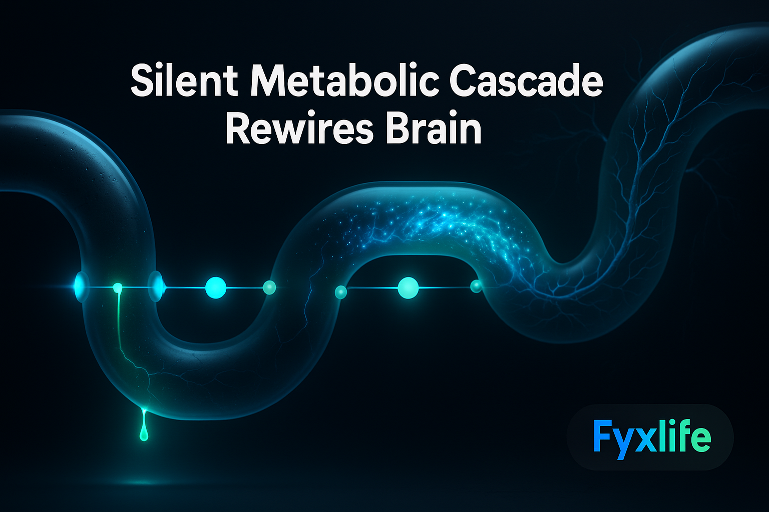
The Three Systems That Feed Each Other — And Feed the Fire
Think of your brain health like a house with three interconnected pipes. When your blood sugar runs chronically high, it’s like acid slowly corroding the first pipe — the one that keeps toxins out of the brain. Once that pipe leaks, pressure floods the second pipe: your brain’s immune system fires up and starts attacking everything in sight, including healthy tissue. That pressure eventually cracks the third pipe — the structural integrity of brain cells themselves. By the time you see water damage on the ceiling — memory loss, brain fog, difficulty concentrating — the corrosion started years ago at the source. The only real fix is upstream, before the first pipe gives way.
These three stages — metabolic damage, chronic neuroinflammation, and structural brain deterioration — do not operate independently. Each one accelerates the next. That compounding is what makes the cascade so dangerous, and so invisible, for so long. It also means that intervening at any single stage has downstream benefits. But the earlier you intervene, the more of the cascade you stop before it starts.
Stage 1 of the Cascade — Metabolic Damage Breaks the Blood-Brain Barrier
How Chronically High Blood Sugar Turns Into Brain Inflammation
Your brain is protected from the bloodstream by a tightly regulated cellular wall called the blood-brain barrier — a selectively permeable lining that controls what enters brain tissue and what gets kept out. Toxins stay out. Nutrients get in. Under normal conditions, it functions like a highly competent border. Under conditions of chronic metabolic stress, it starts to fail.
When blood sugar runs persistently elevated — not diabetically high, but chronically above optimal — the resulting process of glycation (the technical term for glucose molecules bonding to and damaging proteins and cells) degrades the structural integrity of blood vessel walls throughout the body, including those that make up the blood-brain barrier. Once that barrier becomes more permeable than it should be, inflammatory molecules that belong in the periphery begin crossing into brain tissue. The first pipe, in other words, starts to corrode. This cascade entry point — chronically high blood sugar triggering a chain reaction of neuroinflammation — is now a central focus of Alzheimer’s prevention research.
The insidious part is that the blood sugar levels involved are not alarming on a standard report. A fasting glucose of 5.6 or an HbA1c (a three-month average of blood sugar control) creeping upward year on year rarely prompts a serious conversation in a ten-minute GP visit. But it should. These are upstream signals. They are the acid hitting the first pipe.
Why Fat Stored Around Organs Is Particularly Dangerous for Neurons
Not all body fat carries the same neurological risk. Visceral fat — the fat stored deep inside the abdominal cavity, wrapping around organs rather than sitting beneath the skin — is metabolically active in a way that subcutaneous fat is not. It secretes a continuous low-level output of inflammatory signalling molecules (collectively called adipokines), which travel through the bloodstream and contribute directly to the systemic inflammation that eventually reaches the brain.
This means that your waist circumference is not merely a cardiovascular risk marker. It is also a neurological one. Inflammation is identified as a core mechanism through which lifestyle exposures translate into brain damage — and visceral fat is one of its most persistent generators. If your blood sugar is elevated and your visceral fat is high, you are running both processes simultaneously, compounding the rate at which the first pipe corrodes.
Stage 2 of the Cascade — Inflammation Takes Over and Won’t Stop
When Your Brain’s Immune System Becomes the Problem
Your brain has its own specialised immune cells, called microglia. Under normal conditions, they function as the brain’s maintenance crew — clearing debris, pruning old synaptic connections, responding to infection or injury and then standing down. The critical word is then. After a normal immune response, microglia return to a surveillance state. In a brain chronically exposed to metabolic and inflammatory stress, they don’t stand down. They stay activated.
This state of persistent microglial activation — sometimes called neuroinflammation — is where the second pipe takes the hit. What began as a defensive response becomes a destructive one. Activated microglia release their own inflammatory signals, which damage surrounding neurons, degrade synaptic connections (the links between brain cells that allow thought and memory to function), and progressively compromise tissue that was previously healthy. The immune system, originally recruited to protect the brain, becomes one of its primary threats.
This process compounds silently. There are no headaches. No obvious cognitive symptoms. Years can pass in this state while the damage accumulates beneath the threshold of anything you’d notice on a normal day.
The Link Between Systemic Inflammation and Plaque Accumulation
Alzheimer’s disease is associated with the accumulation of two abnormal protein structures inside brain tissue: amyloid plaques (clumps of misfolded protein that build up between neurons) and tau tangles (twisted protein fibres that form inside neurons and disrupt their internal transport system). For decades, these plaques and tangles were treated as the disease itself. The emerging picture is more complicated — and more actionable.
Chronic neuroinflammation appears to accelerate both amyloid accumulation and the spread of tau pathology. The inflammatory environment impairs the brain’s natural clearance mechanisms — including the recently discovered glymphatic system (the brain’s overnight waste-clearance network, most active during deep sleep) — which normally flush amyloid from brain tissue. When that clearance system is compromised, and when inflammation is simultaneously driving protein misfolding, the plaques accumulate faster. The second pipe, under pressure, begins to crack the third.
This is also where the standard health system tends to fall short. The question of whether your specific metabolic profile is quietly feeding neuroinflammation is not one that a routine annual check-up was designed to answer — not because GPs don’t care, but because population-level reference ranges were never built to account for your individual risk profile, family history, or the interaction between your specific biomarkers.
Stage 3 of the Cascade — Structural Brain Deterioration and Grey Matter Loss
What Grey Matter Loss Actually Means for Memory and Focus
Grey matter is the outer layer of the brain containing the actual neuronal cell bodies — the processing centres responsible for memory, decision-making, language, and emotional regulation. Some grey matter loss is a normal part of ageing. The kind that precedes Alzheimer’s is not normal — it is accelerated, region-specific, and measurable years before clinical symptoms appear.
The hippocampus — the brain region most directly involved in forming new memories — is typically among the first areas to show accelerated grey matter loss in people on a trajectory toward Alzheimer’s. When you notice that you’re losing words mid-sentence, or forgetting the name of someone you’ve known for years, or struggling to hold a complex idea in working memory, you are experiencing the downstream output of this structural change. The ceiling has water damage. The pipe broke years ago. New research has found that dietary patterns measurably affect grey matter preservation as the brain ages — which means this stage, too, has upstream levers.
Why Muscle Mass Loss Accelerates This Final Stage
The connection between skeletal muscle and brain health is one of the most underappreciated links in the entire cascade. Muscle tissue is not passive storage. It is metabolically active, secreting its own signalling molecules called myokines — including a protein called BDNF (brain-derived neurotrophic factor), which functions as a kind of fertiliser for neurons, supporting their survival, growth, and connection with each other. When muscle mass declines — as it does naturally from the mid-thirties onward without active resistance training — BDNF output falls with it.
Simultaneously, loss of muscle mass impairs whole-body glucose regulation, worsening the metabolic environment at Stage 1. The cascade feeds back on itself. Physical fitness and resistance training provide greater protective benefit against cognitive decline than brain games and puzzles — not because puzzles are useless, but because muscle mass is doing work that no app can replicate.
Where You Can Break the Cascade
The MIND Diet as a Structural Brake
The MIND diet (short for Mediterranean-DASH Intervention for Neurodegenerative Delay) is a dietary pattern that combines elements of the Mediterranean diet and the DASH diet, specifically optimised for brain health. It emphasises leafy green vegetables, other vegetables, nuts, berries, legumes, whole grains, fish, poultry, olive oil, and a moderate amount of wine — while limiting red meat, butter, cheese, pastries, and fried food. It is not a calorie-restriction protocol. It is a pattern of food choices that systematically reduces the inflammatory and glycaemic inputs that drive Stage 1 of the cascade.
Research has found that the MIND diet helps protect the ageing brain against structural deterioration, specifically grey matter loss. In a region like Southeast Asia, where refined carbohydrate load in daily eating is high and ultra-processed food is increasingly prevalent, this finding is not abstract. Kopi with condensed milk twice a day, white rice at every meal, and routine char kway teow carry a metabolic cost that accumulates over years — and eventually shows up in brain tissue.
Resistance Training: The Most Underrated Brain Intervention
If you are currently spending time on brain-training apps to protect your cognition, you are not wasting your time — but you are spending it on the less effective tool. The evidence for aerobic and resistance exercise as cognitive protection is substantially stronger. Resistance training in particular addresses the cascade at multiple points simultaneously: it improves glucose regulation (Stage 1), reduces systemic inflammation (Stage 2), and increases BDNF output to support neuronal health (Stage 3). Lifestyle interventions — and physical fitness specifically — are identified by researchers as among the most critical factors in protecting brain health against Alzheimer’s progression.
Two to three sessions of progressive resistance training per week is not a gym vanity project. It is, at this point, a neurological intervention. The goal is not aesthetics. It is maintaining the metabolic and myokine-producing machinery that keeps the cascade from accelerating.
Sleep, Toxin Load, and Social Engagement as Cascade Modulators
Deep, uninterrupted sleep is when the brain’s glymphatic system does its clearance work — flushing amyloid and other metabolic waste from brain tissue overnight. Chronic sleep deprivation, or sleep that is disrupted by apnoea or high cortisol, impairs this process directly, accelerating plaque accumulation. Seven to nine hours of quality sleep is not a lifestyle preference. It is a functional maintenance window for the brain.
Environmental and dietary toxins compound inflammatory burden through the same inflammatory pathways that drive the cascade — adding a second upstream entry point that conventional prevention advice often underweights. Minimising chronic exposure to ultra-processed food additives, alcohol, and where possible environmental pollutants reduces the total inflammatory load your brain is managing.
Social engagement, meanwhile, does something that is harder to quantify but consistently appears in the data: it supports cognitive reserve — the brain’s capacity to absorb damage without immediately expressing it as symptoms. Active, meaningful social connection appears to build structural redundancy into neural networks. It is not a soft recommendation. It is a hard one.
What to Track Now — Before You Have Symptoms
The Biomarkers and Lifestyle Variables Worth Monitoring in Your 40s and 50s
Advances in understanding Alzheimer’s biology and biomarkers are actively driving new prevention strategies, with early detection now a viable clinical tool. You do not need to wait for memory symptoms to begin tracking meaningful signals. The metabolic and inflammatory markers that sit at Stage 1 of the cascade are accessible, measurable, and actionable right now.
- Fasting glucose and HbA1c — your primary blood sugar control indicators
- Fasting insulin — a more sensitive early signal of insulin resistance than glucose alone
- hsCRP (high-sensitivity C-reactive protein) — a marker of low-grade systemic inflammation
- Waist circumference — a proxy for visceral fat load
- Grip strength — a validated surrogate for overall muscle mass and physical resilience
- Sleep quality and duration — trackable via wearable or structured self-assessment
- Resting heart rate variability — a measure of nervous system regulation and recovery capacity
None of these require a specialist. Most are available through a standard blood panel. Together, they give you a real-time read on where the cascade currently stands in your own body.
When to Have a Serious Conversation With Your Doctor
If you have a first-degree relative who developed Alzheimer’s or a related dementia, the conversation about biomarker monitoring should happen now — regardless of your current age or symptom status. Genetic risk factors like carrying the APOE-ε4 variant (a specific gene variant associated with significantly increased Alzheimer’s risk) can inform both the urgency and the specific nature of monitoring. Asking your GP whether genetic risk screening is appropriate for your family history is a reasonable and specific question, not an overcautious one.
Beyond genetics, if your metabolic markers are trending in the wrong direction year on year — even within ranges your doctor hasn’t flagged — that trend line matters more than any single reading. Bring the trajectory, not just the number.
This week, check your most recent fasting glucose and HbA1c results. If your fasting glucose is above 5.5 mmol/L or your HbA1c is trending upward — even within the ‘normal’ range — you are looking at Stage 1 of this cascade in motion. That single number is your upstream signal. Bring it to your next doctor visit and ask specifically: “Is my metabolic profile increasing my long-term neuroinflammation risk, and what should I be tracking over the next year?”